

Introduction
I’ll start with a clear definition: tissue dissociation is the controlled process of breaking tissue into individual cells for analysis. In single-cell workflows, tissue dissociation single cell prep sits at the front line of data quality and downstream integrity. Recent audits and bench comparisons report that poor dissociation can cut usable cell yield by 30–60% and reduce viable cell counts dramatically, which raises the stakes for both diagnostics and discovery. So, when a lab tells me they lost half their sample to processing — I ask: where did the protocol fail, and what can we do about it? (I keep a cautious eye on contamination vectors and mechanical stress.) That leads us into the roots of the problem and toward practical choices for better outcomes.
I’ve seen three recurring scenarios: rushed protocols, mismatched enzymes, and over-aggressive mechanical steps. Each one chips away at viability or skews cell representation. The data are blunt — biased populations mean misleading single-cell RNA-seq or poor flow cytometry gating later on. In this article I’ll walk through where traditional approaches fall short, what hidden pains users feel at the bench, and which technical principles can reduce error. We’ll close with concrete metrics you can use to evaluate methods and tools.
Traditional Flaws and Hidden User Pain Points
I’ll be blunt: many labs still rely on decades-old recipes for tissue dissociation, and those recipes were never designed for modern single-cell demands. Directly put — the old ways trade throughput for fragile cell types, and they hide biases. Enzymatic digestion times are often copied without tuning. Mechanical dissociation techniques vary wildly between operators. The result is inconsistent cell viability and unpredictable cell yield. Look, it’s simpler than you think: one minute too long with a protease can wipe out a whole cell subset.
Why does it fail so often?
From my experience, four issues repeat: protocol drift, operator variability, inadequate instrumentation, and poor QC checkpoints. Operator variability creates an attack surface for errors — small differences in pipetting, temperature, or timing accumulate. Protocol drift happens when tweaks are made without recording effects. In enzymatic digestion, you must balance cleavage strength against preserving surface markers. Mechanical dissociation can shear membranes and trigger stress pathways that alter gene expression before you even sequence. These are not theoretical; they show up in lower cell viability, biased population representation, and noisy transcriptomes. I don’t sugarcoat it — it gets messy when teams mix manual tricks with high-throughput goals.
Tools that simply “move faster” often hurt sample integrity. Flow cytometry readouts and downstream microfluidics both suffer when the input suspension is uneven or full of debris. I’ve watched teams chase throughput and lose reproducibility — then scramble to explain why clusters look wrong. In short, traditional methods expose three hidden pains: unpredictable QC, wasted precious samples, and the mental load on technicians who must continually troubleshoot. These pains add real cost — both financial and emotional — to single-cell projects.

New Principles and Practical Outlook
Moving forward, I focus on principled improvements rather than flashy claims. New approaches center on controlled, reproducible steps: calibrated enzymatic mixes, standardized mechanical modules, and inline QC that flags problems early. For example, integrating gentle mechanical dissociation with time-controlled enzymatic digestion reduces cell stress and increases viable yield. I’ve tested workflows where a measured pulse of mechanical agitation plus shorter enzymatic exposure preserved surface markers far better than brute force methods. That matters when you need accurate cell-type proportions later on.
What’s Next
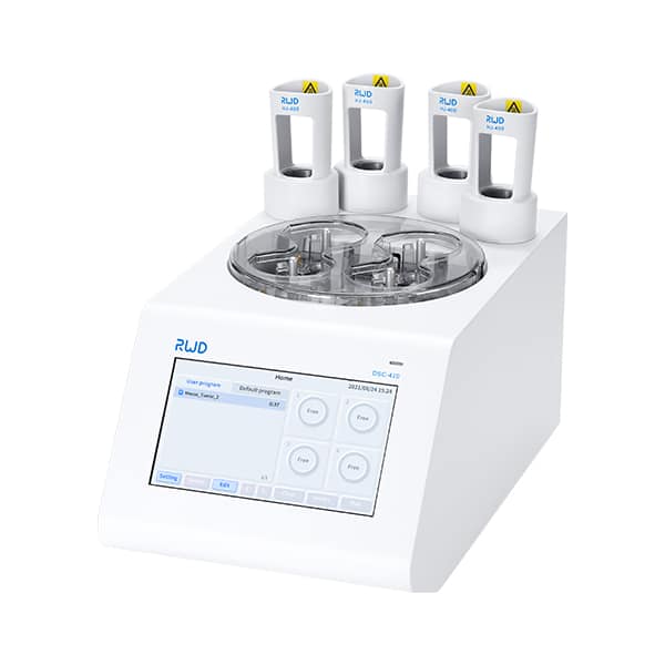
We also see a push toward closed-system dissociators and automated protocols that remove operator variability. The concept is simple: reduce the human error margin and log every parameter. That yields data you can trust (and reproduce across labs) — funny how that works, right? Comparing platforms, I look for systems that allow per-tissue tuning, real-time viability readouts, and easy integration with downstream microfluidics and flow cytometry.
To help teams choose, here are three evaluation metrics I use personally when comparing solutions: 1) Viable cell recovery percentage across replicate samples; 2) Preservation of key surface markers measured by flow cytometry after processing; 3) Protocol traceability — the ability to log and reproduce exact digestion and agitation parameters. If a system scores well on those, it reduces surprises and saves time and samples. I want labs to stop throwing precious specimens at opaque methods and start choosing tools that protect sample integrity. In my view, that shift is the most practical path to better single-cell data.
For labs assessing options now, remember to test on your actual tissue types, not just vendor demos. We’ve seen big differences between tissue classes and a standard protocol. I encourage teams to pilot small batches and compare both yield and transcriptional fidelity before scaling. If you want a calibrated solution that ties into these principles, check leading vendors and read the logs — I trust platforms that prioritize reproducibility over buzz. For more targeted devices and consumables, consider exploring offerings from BPLabLine.